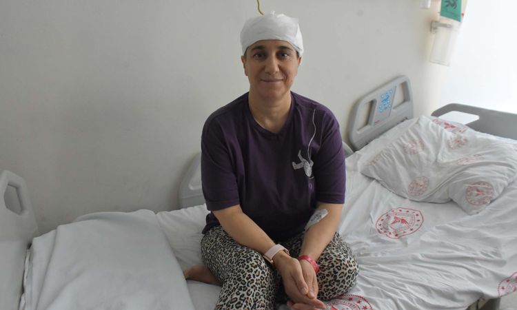
The approximately 8-centimeter tumor in the brain of Songül Bacak (47), a mother of two children living in UŞAK, was successfully removed with the hybrid method applied at Health Sciences University (SBÜ) İzmir Tepecik Training and Research Hospital. In the joint operation of the Interventional Radiology and Brain Surgery teams, the vessels feeding the tumor were first closed by angiography, and then the tumor was completely removed with surgery that lasted approximately 6 hours. Bacak, who regained his health, said, "I feel like I was born again."
Songül Bacak, who lives in Uşak, began to experience short-term fainting, visions, headaches and nausea for the last two years. Bacak, who did not know that the symptoms he was experiencing were seizures, applied to the hospital after his complaints increased. During the examinations, a tumor approximately 8 centimeters in size was detected in Bacak's brain. Bacak, who was treated at SBU Izmir Tepecik Training and Research Hospital, where he was referred, regained his health with the operation performed jointly by the Interventional Radiology and Brain Surgery teams on July 7, with a multidisciplinary approach. Explaining the difficult process she went through, Songül Bacak stated that she was not afraid when she learned that she had a brain tumor and that she had absolute trust in her doctors and said, "I had a headache, nausea and momentary dreams for two years. Turns out these were seizures, I learned when I came here. My doctor told me all the risks at the beginning, but he gave me so much confidence that I went into the surgery without any fear. When I opened my eyes after the surgery, I felt like I was born again. I am very happy. I have two children, one 27 years old and the other 22 years old. "I have been married for 28 years. I love life, my children and my wife. We still have good days to live. I can walk on my own now, I am very well."
Ferhat Bacak (51), who stated that they felt a great relief when his wife regained his health, said, "Of course, we were very scared when we heard that he would have brain surgery, we signed it by risking everything, including the risk of death. We trusted our doctors. Thank God, we survived that trouble, we are very happy now."
FIRST, THE VESSELS OF THE TUMOR WERE CLOSED WITH ANGIOGRAPHY
Interventional Radiology Specialist Dr. Serhat Akis stated that since the tumor was very large, they introduced modern technology to minimize the risk of bleeding during the surgery. Explaining this hybrid method, which is applied in a few centers in Türkiye and the world, Dr. Stream said, "Our patient had a very large tumor of approximately 8 centimeters on his head. Taking lesions of this size directly into surgery carries serious bleeding risks. Therefore, before the surgery, we mapped the brain vessels with the brain angiography method in interventional radiology. Using our advanced angiography devices, we reached only the vessels feeding the tumor without any damage to healthy tissues and blocked these vessels with special closure devices. Thus, in a sense, we prepared the tumor for surgery. This method not only reduces the bleeding in the surgery, but also allows the surgeon to stay alive." "It provides a much safer and more comfortable surgery opportunity by making it easier to distinguish tissue from dead tissue," he said.
THE ENTIRE TUMOR WAS CLEANED IN A 6-HOUR SURGERY
Assoc. Prof. in charge of the Brain and Nerve Surgery Training Clinic, who performed the operation. Dr. Alper Tabanlı stated that the surgery, which took place on July 7 and lasted approximately 6 hours, was completed successfully. Assoc. Dr. Tabanlı continued as follows:
"When our patient arrived, we saw a very large lesion of 8-9 centimeters during the examinations. In the past, we could remove these tumors directly by surgery without these methods, but the risk was extremely high. Working in coordination with our interventional radiologist, we first reduced the blood supply to the tumor. In this way, the surgery time was shortened, the amount of bleeding decreased and the risks of general anesthesia were minimized. During our post-operative checks, we found that the tumor was completely cleared without leaving any residue (recurrence) behind. Our patient is a special patient. "There are other small lesions in his brain. Therefore, genetic research continues. We will follow him closely in the 1st and 6th months."